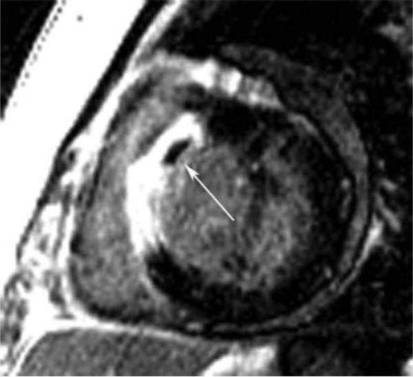
Figure 7.
Microvascular obstruction of a patient after anteroseptal myocardial infarction. This figure is a short-axis late gadolinium-enhanced inversion recovery gradient echo axis image obtained 10 minutes after gadolinium infusion in a patient on Day 3 after reperfused anteroseptal myocardial infarction. Note the transmural late gadolinium enhancement in the anteroseptum. The arrow points to a region of microvascular obstruction in the core of the infarction that represents a region of capillary damage to the extent that contrast is unable to fill this region even 10 minutes after contrast. MO is generally only seen in the first 7 to 10 days post-myocardial infarction and signifies an infarction and patient with poorer prognosis than those without MO. MO indicates microvascular obstruction.

