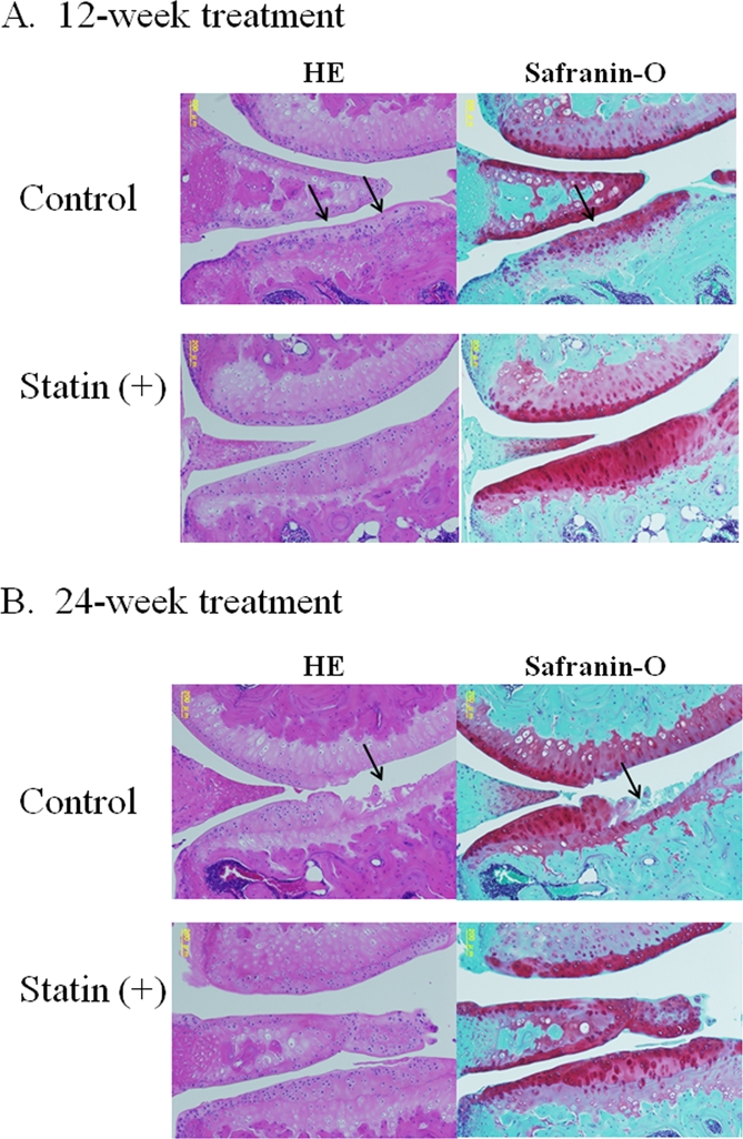
Figure 3. Representative images of cartilage degeneration in OA model mouse.

(A) At 12 weeks, the cartilage in the control group showed a progression of following cartilage degeneration: chondrocyte clustering, abnormal deposition of chondrocytes, and a decreased cell density with cartilage thinning. The cartilage in the statin-treated mice showed less severe in contrast to the control group. (B) After 24-week treatment, most of the control cartilage showed decrease in thickness, marked loss of proteoglycan, chondrocyte clustering, cell death and severe degeneration of cartilage structure, whereas treatment with statin markedly inhibited the degeneration of articular cartilage in the OA mouse model.
