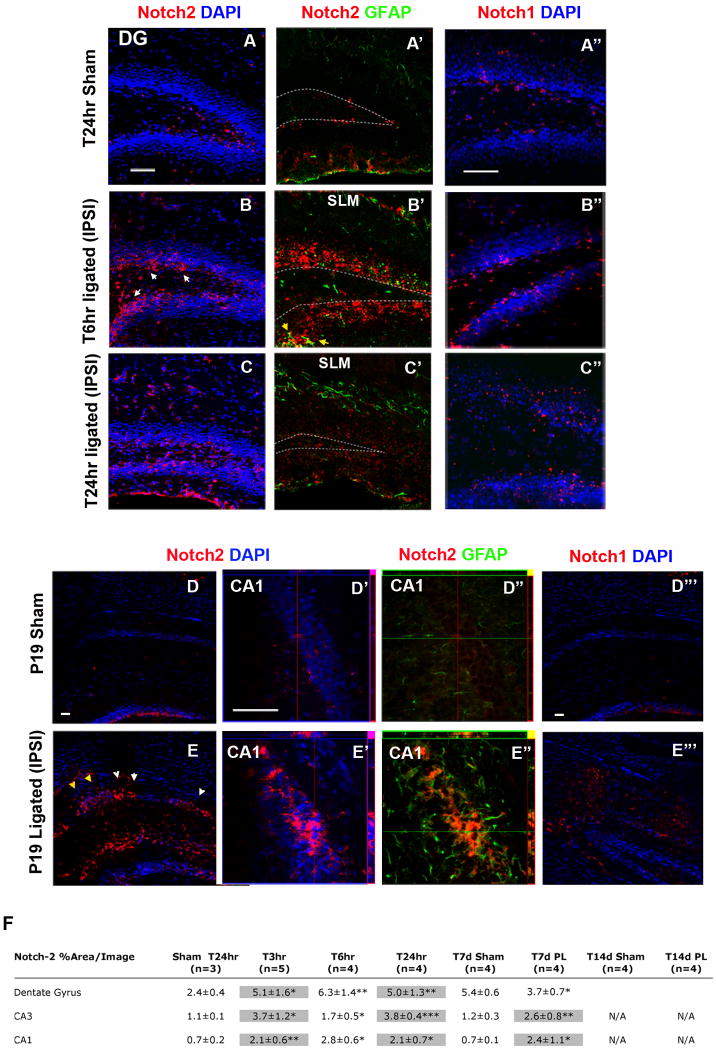
Figure 4. Notch-2 receptor expression and activity in the ipslateral hippocampus, acutely and 7 days following ischemic injury at P12.
(A-A″) Sets of double immunohistochemistry for Notch-2 and Notch-1 show that at T24hr after sham surgery, Notch-2 and Notch-1 expression is restricted to the SGZ. (B-B′) At T6hr after stroke, Notch-2 expression extends to the granule cell layer (GCL) of ipsilateral DG (white arrows). In addition, Notch-2 is expressed only by scattered GFAP+ positive putative astroglia (yellow arrows in B″). (B″) Notch-1 at this time remains restricted to the SGZ. (C-C′) Subsequently, at T24hr after ligation, Notch-2 expression persists ipsilaterally in some cells of the GCL and in the soma of putative astrocytes, invading the stratum lacunosum moleculare (SLM). (C″) Notch-1 expression appears punctuate in the granule cell layer of the DG. (E-E′) 7 days after injury, at P19, Notch-2 is strongly increased as compared to sham control (D-D″) in ectopic niches in the ipsilateral CA1 in and around areas with elevated c-fos expression (yellow arrows, and see Fig. 3, panel A). Orthogonal views of stacked images reveal that Notch-2 is expressed in the granule cell layer in and around condensed nuclei. (E″) Double labeling with Notch-2 and GFAP staining shows that Notch-2 and GFAP expression only modestly co-localize. (E‴) At T7d Notch-1 is only moderately expressed in invading glia and in the granule cell layer. (F) Table summarizing the time course of Notch-2 over-expression in hippocampal region CA1, CA3 and DG. Highlighted boxes indicate regions with elevated apoptosis and c-fos expression. (All scale bars are 50μm; *: p<0.05, ** : p<0.01, *** : p< 0.001).

