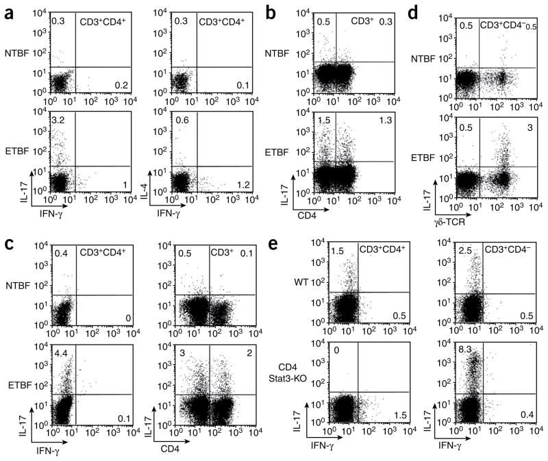
Figure 3.
ETBF, but not NTBF, induces IL-17–producing CD3+CD4+ T lymphocytes and γδ T lymphocytes in the colon lamina propria of Min and WT mice 1 week after NTBF or ETBF inoculation. (a) ICS for IL-17, IFN-γ and IL-4 in CD3+CD4+ T lymphocytes of Min mice. Dot plots are derived from the CD3+CD4+ gate. (b) ICS for IL-17 in CD3+CD4+ and CD3+CD4− lymphocytes from the lamina propria of ETBF-colonized Min mice. Dot plots are derived from CD3+ gate. (c) ICS for IL-17 and IFN-γ in CD3+CD4+ and CD3+CD4− T lymphocytes of C57BL/6 mice. Dot plots are derived from CD3+CD4+ and CD3+ gates. (d) ICS for IL-17 in γδ T cells from the lamina propria of ETBF-colonized Min mice. Dot plots are derived from CD3+CD4− gate. (e) ICS staining in CD3+CD4+ and CD3+CD4− lymphocytes from WT and CD4 Stat3-KO C57BL/6 mice. Dot plots are derived from the CD3+ gate. Each panel is representative of at least three independent experiments except e (two independent experiments). The numbers inside the plots indicate the percentage of the cell population showing the quadrant characteristic.

