
FIGURE 1.
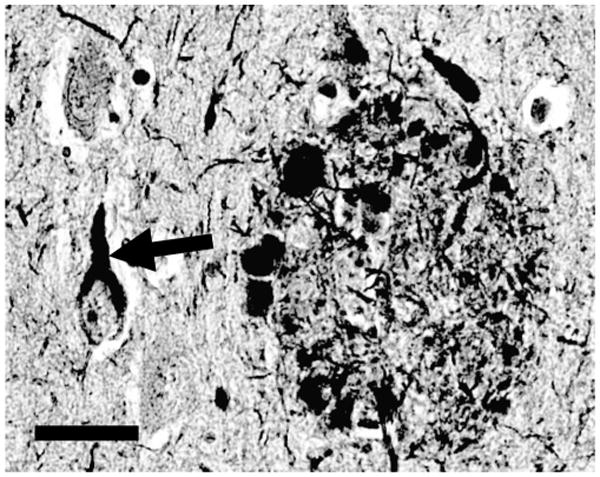
Histopathologic hallmarks of Alzheimer disease (AD). A neurofibrillary tangle (diagonal arrows) and neuritic plaque (oval-shaped structure on right) are shown in a photomicrograph from human AD brain section stained with the silver-impregnation Bielschowsky technique. Neurofibrillary tangles are composed of insoluble and protease-resistant fibrils, and develop intracellularly. Neuritic plaques are extracellular fibrillary amyloid deposits, surrounded by swollen, degenerating, silver-impregnated neurites. Scale bar = 50 μm.