
Figure 6.
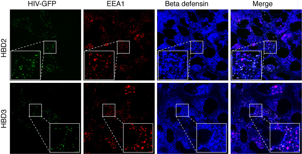
Localization of HIV in the HBD2 and HBD3 containing early endosomal vesicles. GFP-labeled HIV-1 81-A virus was added to the apical surfaces of polarized adult tonsil epithelial cells. Cells were incubated at 37°C for 2 h. Cells were then fixed and co-immunostained for EEA1 (red) and HBD2 or HBD3 (both in blue). Cells were analyzed by confocal microscopy and images were taken about 3 µm below the apical surface. The pink in merged panels indicates co-localization of EEA1 (red) with HBD2 or HBD3 (blue). The white in the merged panels indicates co-localization of HIV (green), HBD2 (blue), and EEA-1 (red), indicating co-localization of HBD2 and virions in the EEA1-positive early endosomes (red).