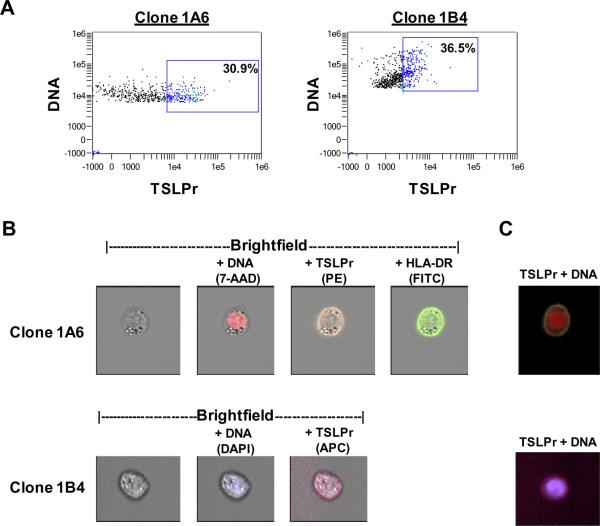
Figure 1. Validation of Anti-TSLP Receptor Monoclonal Antibodies by Image-Based Flow Cytometry.
Activated THP-1 monocytes were stained for TSLPr, HLA-DR and DNA. (A) Dot plots representing gated focused single cells with DNA negative cells excluded. Boxes denote TSLPr+ gates determined by visual inspection of single cells inside the gate versus cells outside the gate. Green crosshairs denote single cells shown in panels B and C. (B) Single cell images of TSLPr+ cells using different anti-TSLPr clones. Images for each stain are overlayed on the brightfield image. (C) Composite images corresponding to cells shown in (B).

