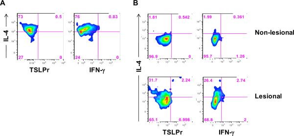
Figure 8. Analysis of TSLPr on Skin T Cells.
(A) Cells isolated from normal skin obtained from a non-atopic donor were cultured for 7 days with TSLP+allergen and analyzed by standard flow cytometry. (B) Cells isolated directly ex vivo from lesional or non-lesional skin from an AD patient (#1) were analyzed for TSLPr. Both panels show density plots for cells in the CD3+ T lymphocyte gate.

