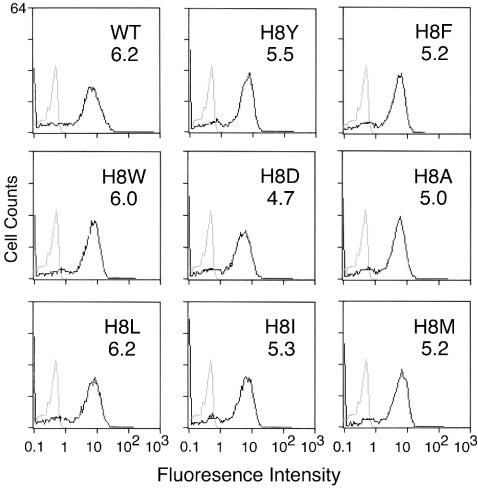
FIG. 2.
Virus binding. Virus supernatant concentrated to 15- to 20-fold by using Centricon Plus-80 (100-kDa cutoff) to remove shedded SU was incubated at 4°C with human 293 cells stably expressing the exogenous ecotropic receptor. Cells were then stained with goat anti-SU antisera (anti-Rauscher gp70; Quality Biotech, Inc.) and mouse anti-goat antisera conjugated to fluorescein isothiocyanate. The level of binding was analyzed by flow cytometry. Gray lines represent the basal level of fluorescence determined by incubation of WT virus with parent human 293 cells lacking ecotropic receptor. For comparison these basal values are shown in each panel. Black lines represent the fluorescence intensity of binding of WT or mutant virus to human 293 cells expressing ecotropic receptor. The value of the mean fluorescence intensity is shown in upper right corner of each panel. Values shown are from a representative of at least two independent binding experiments.

