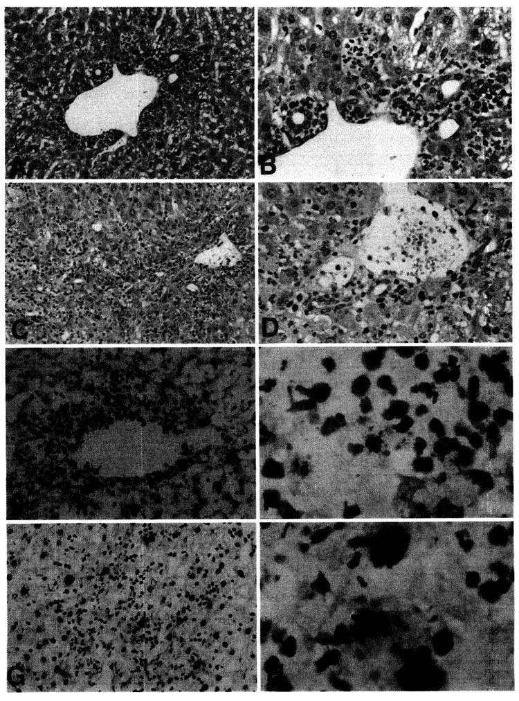
Figure 6.

(A–D) Histological appearance of FL-treated B10 donor liver allografts 4 days after transplantation into normal C3H recipients. (A and B) normal liver allograft, showing predominantly periportal inflammation (A), with absence of damage to bile duct epithelium (B). (C and D) FL-treated donor liver allograft, showing diffuse periportal and lobular inflammatory cell infiltration, with focal bile duct injury (arrowheads) and evidence of endothelial cell separation from a portal vein (arrow). (Hematoxylin and eosin stain; A and C, original magnification, ×100; B and D, original magnification, ×200.) (E–H) TUNEL staining for apoptotic cells in liver allografts, 4 days after transplantation. (E and F) normal liver allograft, showing apoptotic cells within the periportal inflammatory cell infiltrate (compare with A). (G and H) FL-treated donor liver allograft, showing larger, TUNEL-positive cells (presumptive apoptotic hepatocytes) throughout the lobular area. (Counterstained with hematoxylin; E and G, original magnification, ×100; F and H, original magnification, ×400.)
