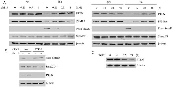
Figure 1. DhS1P reverses constitutive phosphorylation of Smad3 in SSc fibroblasts through up-regulation of PTEN/PPM1A protein levels.
A. Healthy (NS) and SSc fibroblasts were treated with increasing doses of dhS1P for 24 hours (left panel) or were treated with 0.5 μM of dhS1P for the indicated time points (right panel). PTEN, PPM1A, Phospho-Smad3, and total Smad3 were analyzed by western blotting. β-actin was used as loading control. B. SSc fibroblasts were treated with PTEN or non-silencing siRNA for 24 hours and stimulated with dhS1P for additional 24 hours. Phospho-Smad3, total Smad3, PTEN and β-actin were analyzed by western blotting. C. Healthy control fibroblasts were treated with TGF-β (2.5 ng/ml) for the indicated time points. PTEN and β-actin levels were analyzed by western blotting.

