
Figure 7. Partial colocalization of FBXO25 with β-actin in nucleus of HeLa cells.
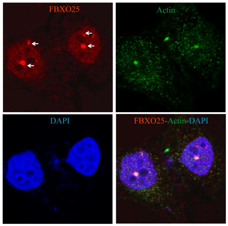
Confocal microscopy of cells double-stained with affinity-purified anti-FBXO25 antibodies and antibodies against β-actin. The arrow points to a FAND. The proteins labeled in each panel are indicated in the upper left and right of the panel in the relevant color. DAPI was used to stain nuclei.