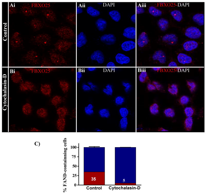
Figure 8. Inhibition of actin polymerization using cytochalasin-D disrupts FANDs.

Confocal microscopy of labeled FBXO25 in untreated and cytochalasin-D-treated (5 μg/ml for 24 h) HeLa cells are shown. Without cytochalasin-D treatment, FANDs were found in the nucleoplasm (Ai–Aiii). In the presence of cytochalasin-D, the majority of endogenous (Bi–Biii) FANDs disappeared (B). (C) Proportion of cells containing FANDs was estimated. After treatment with cytochalasin-D, the proportion of cells containing FANDs (red) significantly differs from the control population. Four separate experiments were performed, and around 100 cells per slides were analyzed for cytochalasin-D treatment.
