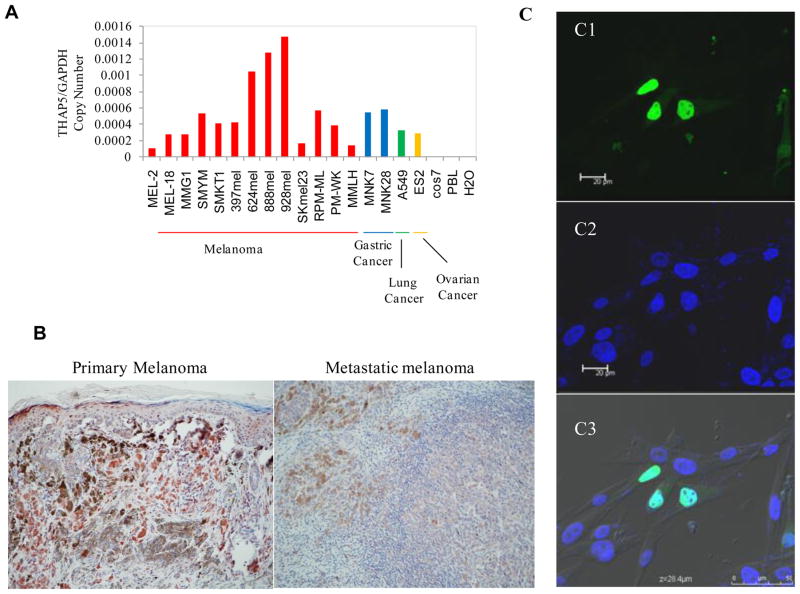
Fig. 1.
Expression and localization of THAP5 in melanoma cells. A, THAP5 mRNA expression in various cell lines. THAP5 is expressed at various levels in all human melanoma cell lines tested. THAP5 expression was also detected in some human gastric, lung and ovarian cell lines but not in Cos7 or PBL cells. B, Imunohistochemistry of primary and metastatic melanoma tissues showing distinct THAP5 staining of melanocytes. C, Subcellular localization of the GFP-THAP5 in MeWo cells. Confocal images of MeWo cells transfected with GFP-THAP51-395 shows nuclear localization (green in C1). Panel C2 shows cells with DAPI staining for the nucleus and C3 shows merged image of Panels C1, C2 and C3.

