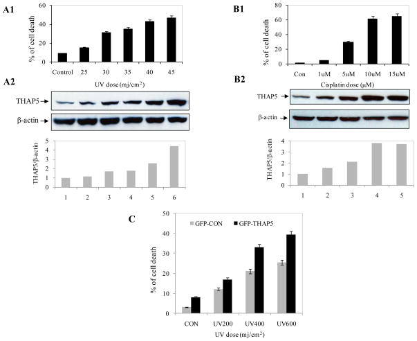
Fig. 2.
THAP5 is induced following UV or cisplatin treatment. MeWo cells were treated with increasing doses of UV and cisplatin and apoptosis monitored by flow cytometry as described in the Methods (A1, B1). Extracts were prepared from the same cell populations and subjected to SDS-PAGE and Western blot analysis using THAP5 antibody. THAP5 was significantly induced with increasing doses of UV and cisplatin and this corresponds to an increased apoptosis in the treated cells (A2, B2). β-actin antibody was used to verify that equal amount of protein was present in each lane. Bottom panels, A2 and B2 show densitometry analysis. C. MeWo cells were transfected with GFPC vector and GFPC-THAP5 plasmids. Transfected cells were exposed to UV and cell death was monitored. Data are mean ± SD of 3 different experiments.

