Figure 1.

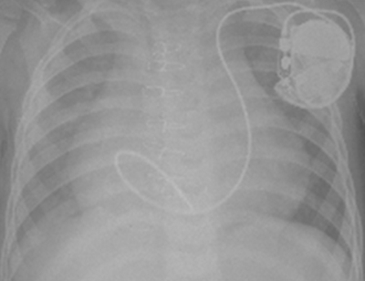
Chest X-ray shows transvenous active ventricular lead implantation via the persistent left superior vena cava and coronary sinus

Chest X-ray shows transvenous active ventricular lead implantation via the persistent left superior vena cava and coronary sinus