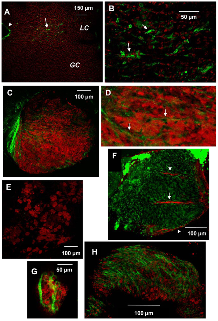
Figure 4.
SMC are positioned to influence lesion development within the LCAA of ApoE KO mice. Shown are en face confocal images of aortic arch tissue from ApoE KO mice of different ages stained with FITC-labeled anti-smooth muscle α-actin (SMA). A, B: SMC (green) are found within the lesion-prone LCAA (LC) of 16 week-old ApoE KO mice but not in the lesion-resistant greater curvature (GC). Cell nuclei are stained with Topro3 (red); arrowhead indicates SMA staining at cut edge of tissue (SMA antibody penetrates into the media at the cut edges only). B is higher magnification of LC in image A. C–G: Lesions of 25 week-old ApoE KO mice were composed of clusters of lipid-laden foam cells. Neutral lipid was visualized with Oil Red O (red) except in F, in which neutral lipid was visualized with BODIPY (green) and mouse anti-SMA was detected with Cy5-conjugated anti-mouse IgG (red). SMC were found intimately associated with some (C,D,F,G), but not all foam cell aggregates (E). C, D, F: Spindle-shaped SMA+ cells surround (arrowhead) and infiltrate (arrows) a large aggregate of foam cells within ApoE KO LC. F: Spindle-shaped cells adjacent to SMA+ cells contain neutral lipid. G: A few SMC envelop a small cluster of few foam cells within TLR4 DKO LC. H: A contiguous SMC layer covers a later lesion in an older ApoE KO mouse (44 wk). C and F are also presented as online supplemental data, consisting of sequential z-plane images, proceeding from the luminal to medial aspect of the lesion.

