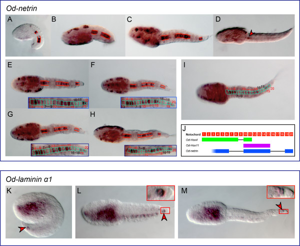
Figure 5.
Uneven expression of Oikopleura netrin and laminin α1 in the notochord. (A-I) Microphotographs of Oikopleura embryos at the mid-tailbud/early hatchling (A), late tailbud (B), Stage 1 (C,E-I), and Stage 2 (D), hybridized in situ with a digoxigenin-labeled Od-netrin antisense probe. All embryos are oriented with anterior to the left. Red lines delineate the notochord domains of Od-netrin expression. Individual notochord cells are outlined in aqua in (I) and in the insets showing the magnified notochord (E-H). In (D), a red arrowhead indicates residual staining in one of the notochord cells close to the trunk-tail boundary. (J) Schematic representation of the Oikopleura notochord cells (20 numbered red squares), followed, for comparison purposes, by a delineation of the Od-Hox4 (green), Od-Hox11 (purple) [29], and Od-netrin (blue) expression patterns. (K-M) Microphotographs of Oikopleura embryos at the mid-tailbud/early hatchling (K), Stage 1 (L), and Stage 2 (M), hybridized in situ with a digoxigenin-labeled Od-laminin α1 antisense probe. A red arrowhead indicates the staining in the terminal notochord cell, which is magnified in the insets in panels (L) and (M).

