
Figure 1.
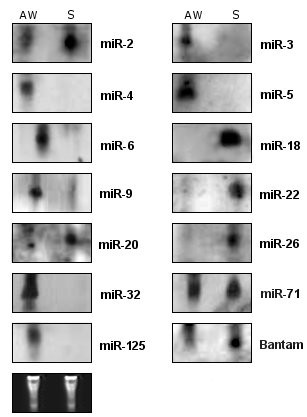
Northern blot analysis of selected miRNAs in two different developmental stages in S. mansoni. Sixty micrograms of total RNA for each sample were separated on a 15% denaturing polyacrilamide gel, blotted and probed using miRNA specific DIG-labeled probes. Lanes from left to right: S. mansoni adult worm pairs (AW) and schistosomula (S) (7 days after mechanical transformation). miR-71, miR-125 and Bantam are the miRNAs identified in S. mansoni homolog to miRNAs of S. japonicum [38]. The tRNA and 5S rRNA bands were visualized by ethidium bromide staining of polyacrylamide gels and served as loading controls and are shown at the bottom.