
Figure 2. Euphol is a bioisoster of MGL.
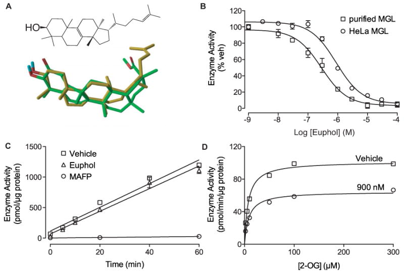
(A) Chemical structure of euphol (top) and 3-dimensional superposition of euphol (gold) and pristimerin (green). Oxygens, red; hydrogens, blue.
(B) Concentration-dependent inhibition of purified MGL (squares) and HeLa MGL (circles) by euphol. Enzyme activity is reported as percent of vehicle controls (DMSO, 1%). Results are expressed as mean ± SEM (n=3).
(C) Rapid dilution assays of HeLa-MGL in the presence of vehicle (squares, DMSO, final concentration 2%), euphol (triangles), or MAFP (circles). We measured the amount of reaction product (pmol/μg protein) generated over a 60-min period following a 20-min preincubation with inhibitor or vehicle. Results are expressed as mean ± SEM (n=3).
(D) Michaelis-Menten analysis of the MGL reaction in the presence of vehicle (squares, DMSO, 1%) (n=4) or euphol (900 nM). Results are expressed as mean ± SEM (n=3).