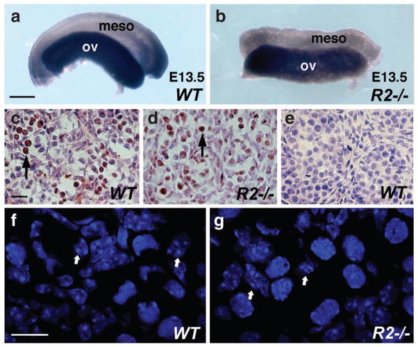
Figure 2. Germ cells in Raldh2 mutant ovary undergo meiosis.
(a, b) Expression of Scp3 in E13.5 wild-type and Raldh2−/− ovaries examined by whole-mount in situ hybridization; scale bar, 200 μm. (c–e) Immunostaining for γ-H2AX in E13.5 ovary (c, d) and testis (e); arrows indicate female germ cells positive for γ-H2AX detection; scale bar, 10 μm. (f, g). Confocal images of nuclei stained with DAPI (4′,6-diamidino-2-phenylindole) in E14.5 wild-type and Raldh2−/− ovaries; arrows indicate representative germs cells in meiotic prophase; scale bar, 10 μm. Meso, mesonephros; ov, ovary. n = 3 for each specimen.

