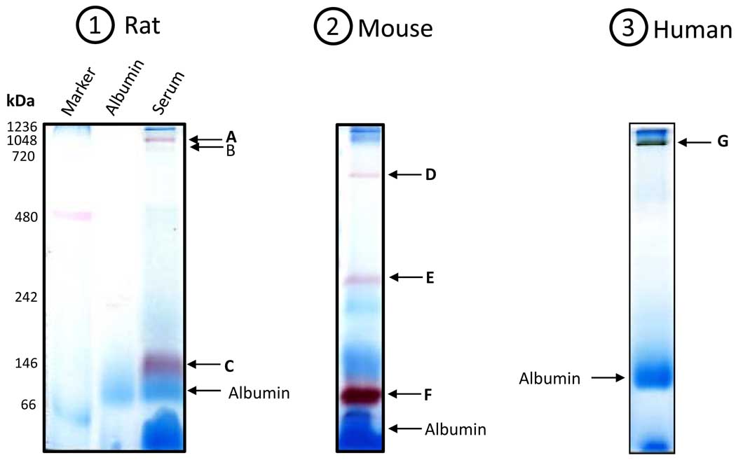
Figure 3.
In-gel staining of esterase activities in the serum from (1) rat; (2) mouse; and (3) human. For rat and mouse serum, β-naphthylacetate was used; for human serum, α-naphthylacetate was used. Gels were incubated in 100 ml 50 mM Tris-HCl, pH 7.4 containing 50 mg naphthylacetate in 1 ml ethanol and 50 mg Fast blue BB salt. Arrows indicate the bands that exhibited esterase activities (A to G). Note that serum from a young rat (3 months old) and a young mouse (5 months old) was used, respectively, for experiments in this figure. All gels were 12% resolving and 4% stacking.

