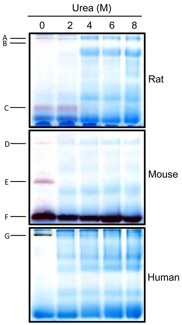
Figure 5.
In-gel detection of serum esterase activity using samples that were treated with varying concentrations of urea. For each species, the serum was diluted to 1 mg/ml in micro-tubes that contained the indicated urea concentrations. Following mixing with BN-PAGE loading buffer, the samples were loaded onto the gels and electrophorized. Nongradient BN-PAGE (all 12% resolving gels) and esterase activity staining were performed as described in Figs. 1 and 3.

