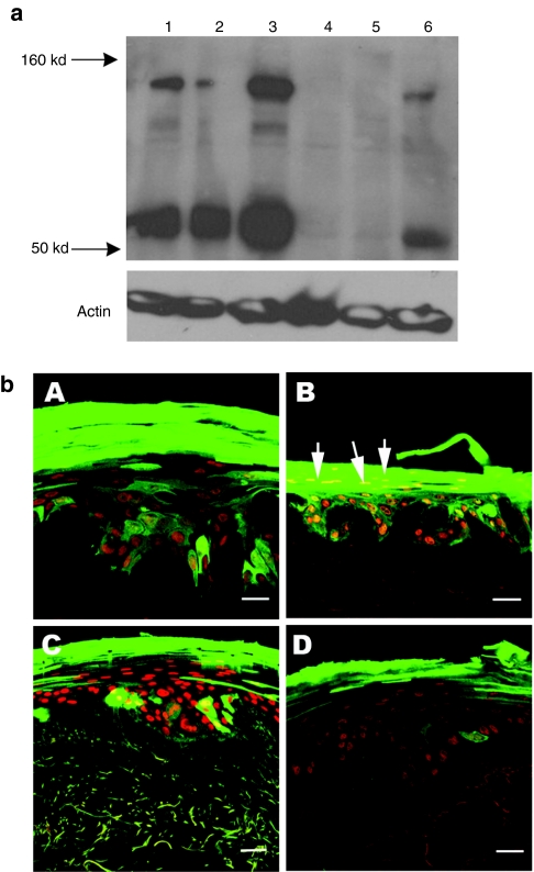
Figure 4.
Lentiviral vector delivery of SPINK5 transgene into primary keratinocytes. (a) Transduced healthy donor or NS primary keratinocytes were harvested and the LEKTI expression was assessed using anti-LEKTI antibody by western blot analysis. Lane 1 (healthy donor) and lane 4 (NS) were nontransduced cells. Endogenous full length (145 kd) and cleaved (~68 kd) LEKTI could be detected in normal keratinocytes nontransduced or transduced with eGFP alone expressing vector (lane 1 and 2), but not in NS cells (lanes 4 and 5). In contrast, in cells transduced with SPINK5/eGFP vector, increased expression of LEKTI was observed in both healthy donor (lane 3) and NS cells (lane 6) compared to the cells transduced with eGFP alone (lanes 2 and 5). These results demonstrate successful reconstitution of LEKTI expression in NS keratinocytes following lentiviral transduction. (b) Primary keratinocytes from health donors or NS were transduced with lentiviral vector expressing SPINK5/eGFP or eGFP alone. GFP+ cells were sorted by flow cytometry to enrich eGFP+ cells and then deployed in organotypic cultures (OTC). eGFP in frozen sections from (OTC) was directly detected under fluorescence microscopy. (A,C) Cultures derived from healthy donor keratinocytes, and (B,D) cultures derived from NS keratinocytes. Cells transduced with eGFP alone (A,B) or SPINK5/eGFP (C,D) all expressed high levels of eGFP (green) with an intense staining pattern especially in the uppermost layers of the epidermis. NS keratinocytes transduced with SPINK5/eGFP showed reduced numbers of nuclei in the cornified layer of the epidermis (D) compared to the culture generated by NS cells transduced with eGFP alone (B, arrowed). Red shows nuclei staining by propidium iodide. Bar = 40 µm. eGFP, enhanced green fluorescent protein; LEKTI, lymphoepithelial Kazal-type-related inhibitor; NS, Netherton syndrome.

