Figure 6.
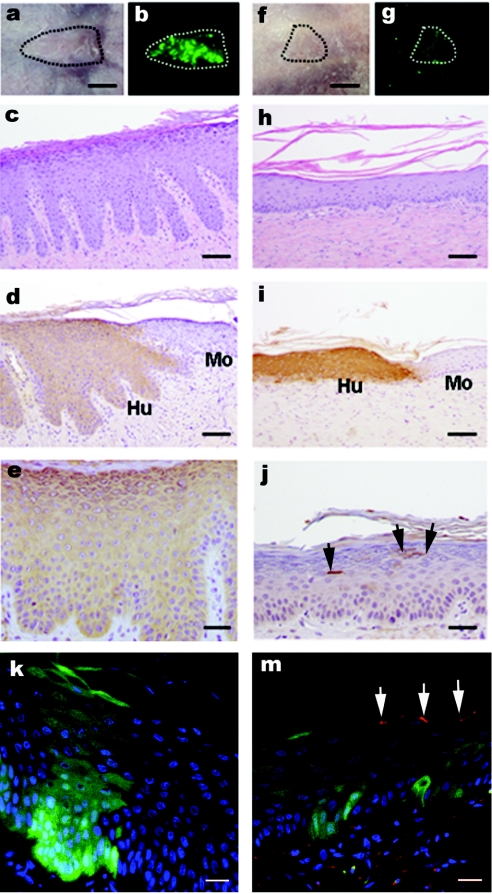
In vivo assessment of NS keratinocyte correction in a humanized mouse model: macroscopic and histological examination following lentiviral transduction. NS keratinocytes were transduced with (a–e,k) eGFP or (f–j,m) SPINK5/eGFP and grafted onto nude mice in two independent series of experiments. Regenerated skin grafts were examined 8 weeks after grafting. (a,f) macroscopic appearance of the graft under transmit light and (b,g) a real-time eGFP expression under 488 nm light. (c,h) Histological appearance of grafts. (d,i) Human involucrin expression using antihuman involucrin antibody to indicate mouse (Mo)—human (Hu) skin boundary. (e) eGFP expression using GFP antibody. (j) The LEKTI expression using antihuman LEKTI antibody and arrows indicate isolated or clustered LEKTI+ cells within a wider area of skin. (k,m) Both frozen sections represent the overlapping between real-time eGFP (green) expression and LEKTI expression (red) stained with LEKTI antibody. These images showed that there was reversal of papillary changes in the graft transduced with SPINK5/eGFP (h) compared to the graft transduced with control vector eGFP (c). Low numbers of LEKTI expression cells in the grafts transduced with SPINK5/eGFP vector in both paraffin and frozen sections (j and m, arrowed areas) suggest small population of corrected cells sufficient to mediate wider correction of the epidermal architecture. bars: (a,b,f,g) = 5 µm; (c,d,h,i) = 200 µm; (e,j) = 100 µm; (k,m) = 40 µm. eGFP, enhanced green fluorescent protein; NS, Netherton syndrome.

