Figure 5.
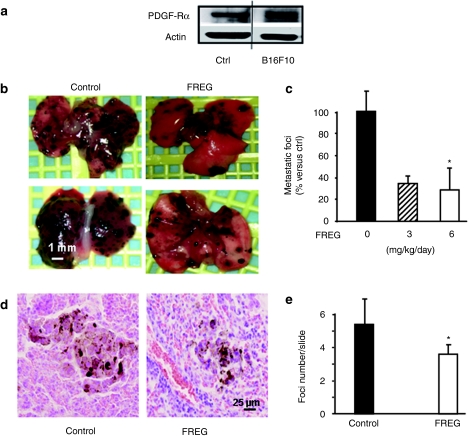
FREG intravenous (i.v.) treatment inhibited melanoma lung metastases in a murine melanoma model. (a) In vitro expression of PDGF-Rα was investigated in growing B16F10 mouse melanoma cell line, by western blotting analysis. Human HUVEC cells were used as positive control, according to previously reported data.26 (b) B16F10 melanoma cells (2 × 10⁵/200 µl PBS) were injected i.v. into C57BL/6 mice tail vein at day −6. Animals were treated i.v. with 50 µl of FREG (3 or 6 mg/kg/day) on days 0, 2, 4, 7, and 8. Macroscopic lung metastases are indicated in representative lungs. Bar = 1 mm. (c) Quantification of macroscopic lung metastases indicates a significant reduction upon FREG treatment. Asterisk (*) indicates statistical significance (P < 0.05 versus ctrl) according to one-way analysis of variance test followed by Dunnett's multiple comparison test. Linear trend post hoc analysis indicates a significant dose-related effect (P < 0.02). (d) Microscopic metastatic foci upon histological analysis. Representative image are reported, indicating representative foci-size. Bar = 25 µm. (e) Quantification of number of microscopic foci per slide indicates a significant reduction upon FREG treatment (P < 0.05 versus ctrl). ctrl, control; PDGF-Rα, platelet-derived growth factor-receptor-α.

