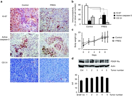
Figure 6.
Immunohistochemical analyses were carried out on mice injected intravenously with B16F10 melanoma cells and treated with FREG (6 mg/kg) at days 0, 2, 4, 7, and 8. (a) The routine hematoxylin–eosin staining and Ki-67, active caspase-3, and CD 31 labeling were performed on paraffin-embedded lungs. Bar = 25 µm. (b) Quantification of percentage of immunoreactivity expression indicates a significant reduction of cell proliferation (Ki-67 staining), a marked induction of apoptosis (caspase-3 activation), and a significant decrease of angiogenesis (CD 31 staining) upon FREG treatment (*P < 0.05 FREG versus control). (c) Total body weight was recorded in healthy C57BL/6 mice at days 0, 2, 4, 7, 8, and 9 in mice treated with 6 mg/kg/day FREG intravenously. No significant differences versus control were observed at any day indicating no gross acute toxicity upon FREG treatment. (d) PDGF-Rα expression was detected by western blotting (top) and by quantitative real-time PCR analysis (bottom) in 5 mouse primary melanomas (obtained by s.c. injection of B16F10 melanoma cells in C57BL6 male mice). The experiments were performed in triplicate; data are reported as average and SD. PDGF-Rα, platelet-derived growth factor-receptor-α.

