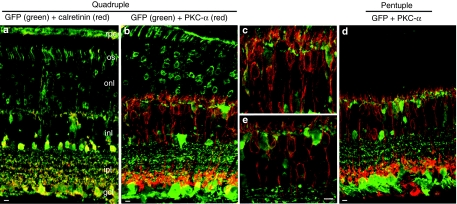
Figure 7.
Evaluation of colocalization of GFP (green) with cell-marker proteins (red), as shown by immunohistochemistry of frozen retinal sections at 1 month following intravitreal injections with different AAV2 Y-F mutant vectors in adult mice. (a) Colocalization (yellow) of GFP and some calretinin (red) positive ganglion or amacrine cells after injection of the quadruple mutant. (b,d) Colocalization of GFP (green) and PKC-α (red) positive rod bipolar in the eyes treated with the quadruple or pentuple mutant vector, respectively; (c,e) Insets of panels (b) and (d) at higher magnification showing colocalization of GFP and PKC-α in the inner nuclear layer. Bar = 10 µm (a;b;d) and 5 µm (c;e). AAV2, adeno-associated virus serotype 2; gcl, ganglion cell layer; GFP, green fluorescent protein; ipl, inner plexiform layer; inl, inner nuclear layer; onl, outer nuclear layer; os, outer segments; rpe, retinal pigmented epithelial layer.

