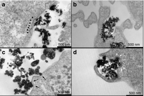
Figure 1.
Uptake of nanoparticles by hCMEC/D3 cells. hCMEC/D3 cells were incubated with NBs, PrPBs, and PEIBs, and investigated with electron microscopy. (a) NBs are found interacting with electron-dense plasma membrane (arrowheads) or (b) within cellular extensions. (c) PrPBs show binding to less pronounced electron-dense regions at the plasma membrane (arrows). (d) Cellular protrusions embrace PEIBs. Structures resembling clathrin-coated pits are indicated with white arrowheads. NB, noncoated bead; PEIB, polyethyleneimine-coated bead; PrPB, prion-coated bead.

