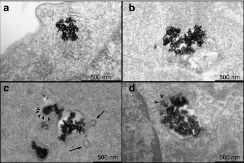
Figure 4.
NBs, PrPBs, and PEIBs localize in morphologically distinct intracellular vesicles. hCMEC/D3 cells were incubated with nanoparticles for 1 hour and 30 minutes. (a,b) NBs were found in vacuoles resembling multivesicular bodies (MVBs) and in multilamellar bodies. (c) PrPBs were primarily localized within vesicular structures. Note the presence of clathrin-coated buds (arrows) and the formation of clathrin latices around the prion coat (arrowheads). (d) PEIBs resided in large endosomes. Clathrin latices were found around the PEI coat (arrowheads). Bar = 500 nm. NB, noncoated bead; PEIB, polyethyleneimine-coated bead; PrPB, prion-coated bead.

