Figure 3.
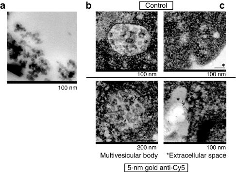
Ultrastructural detection of miR-143 in the MV-rich fraction. (a) MVs observed by electron microscopy in the MV-rich fraction. (b,c) Immunoelectron microscopic study. Immunogold-anti-Cy5 was used for the detection of Cy5, with which miR-143BP had been labeled. The MVs in the (b) multivesicular body and (c) extracellular space are shown, and the specific signals (gold particles) indicating miR-143BP/Cy5 are indicated by the arrows. There is no signal in the controls (upper photos). MV, microvesicle.

