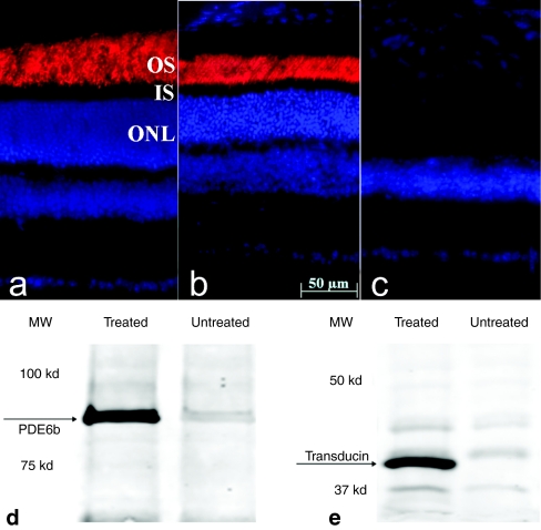
Figure 5.
PDEβ expression in treated and untreated eyes of rd10 mice. PDEβ immunostaining (red) in a 6.5-month old (a) uninjected normal C57BL/6J eye, (b) treated, and (c) untreated eye from one rd10 mouse. Nuclei were stained with DAPI (blue). Note the robust staining of PDEβ in outer segments of the treated rd10 eye compared to its absence in untreated, contralateral control eye. Western blot showing (d) abundant PDEβ and (e) rod transducin-α expression in treated but not in untreated rd10 eyes. PDEβ, β subunit of rod cGMP-phosphodiesterase; OS, outer segments; IS, inner segments; ONL, outer nuclear layer.

