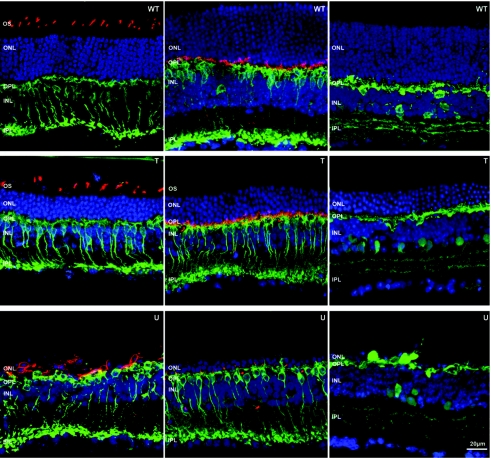
Figure 6.
Comparisons of retinal morphology in normal (WT), P14 + 6M treated (T) and untreated (U) rd10 retinas. Blue: nuclear counterstaining with TOTO-3. Left column, green: rod bipolar cell specific Protein kinase C α (PKC α) Ab staining; red: S- and M-cone opsins Ab staining. Middle, green: bipolar cell specific PKC α Ab staining; red: postsynaptic density protein 95 (PSD95) that stains PR synaptic terminals in the outer plexiform layer (OPL). Right, green: horizontal and amacrine cell specific Calbindin Ab staining. PR, photoreceptor; OS, outer segments; ONL, outer nuclear layer; INL, inner nuclear layer, IPL, inner pexiform layer, WT; wild type.

