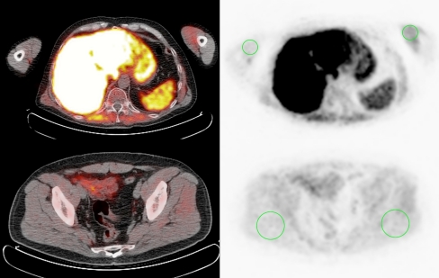
Fig. 1.
Muscle uptake quantification. Transverse slices of [18F]methylcholine PET (right) and image fusion with low-dose CT (left), showing the regions of interest used for evaluation of muscle uptake (green circles). Volumes with a thickness of 5 slices were delineated in the biceps (top) and gluteus maximus (bottom) muscles on both sides

