Figure 6.
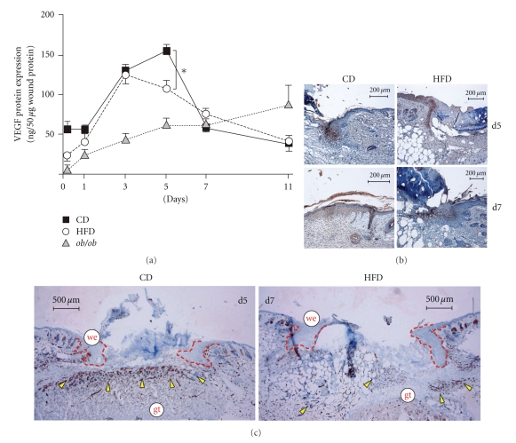
Angiogenic processes at the wound site. (a) VEGF ELISA analysis from lysates of nonwounded back skin and lysates of wound tissue isolated from CD, HFD or ob/ob mice. VEGF protein is expressed as ng per 50 μg skin or wound lysate. *P < .05 as indicated by the bracket. Each single experimental time point depicts the mean ± SD obtained from 8 wounds (n = 8) isolated from 4 individual animals (n = 4). Sections from 5-day and 7-day wound tissue isolated from CD and HFD mice (as indicated) were stained for VEGF (b) or CD31 (c) Protein (brown colour). The epithelial margins are indicated by a red line. Scale bar equals 200 μm in (b) or 500 μm in (c). gt, granulation tissue; we, wound margin epithelia.

