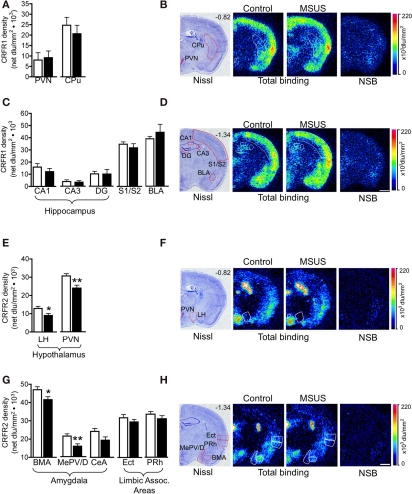
Figure 8.
Corticotropin releasing factor receptor binding in F1 MSUS and control mice. (A,C) Normal CRFR1 binding was observed in several brain areas in F1 MSUS brain (MSUS, n = 5–6, control, n = 5–6). (E,G) Reduced CRFR2 binding was observed in several hypothalamic and amygdala subnuclei, but not in cortical areas in F1 MSUS brain (MSUS, n = 12, control, n = 6–7). (B,D,F,H)Sections showing Nissl staining (B,D) total CRFR1 binding and CRFR1 non-specific binding (NSB), and (F,H) total CRFR2 binding and CRFR2 NSB, in different brain areas in F1 control and MSUS mice. Number in top right corner of Nissl section indicates Bregma position. Scale bar 1 mm. *p < 0.05; **p < 0.01. BLA, basolateral amygdala; BMA, basomedial amygdala; CPu, caudate putamen; DG, dentate gyrus; Ect, ectorhinal cortex; LH, lateral hypothalamus; PVN, paraventricular nucleus; MePV/D, medial posteroventral and medial posterodorsal amygdala; PRh, perirhinal cortex; S1/S2, somatosensory cortex. Bar graphs represent mean values ± SEM.

