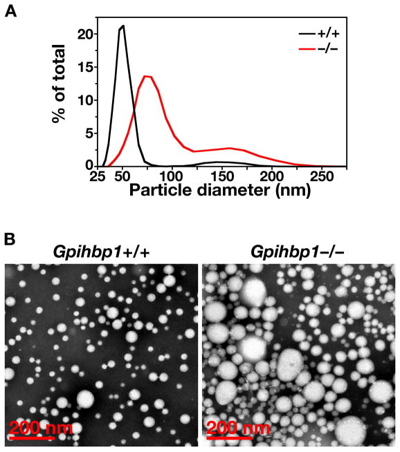
Figure 17.
Distribution of lipoprotein diameters in the d < 1.022 g/ml lipoproteins from Gpihbp1−/− and Gpihbp1+/+ mice. (A) As judged by dynamic laser light scattering, the median diameter of lipoproteins was 157% larger in Gpihbp1−/− mice (n = 3) than in Gpihbp1+/+ mice (n = 6). 15.4% of the particles in Gpihbp1−/− mice had diameters of 122–289 nm. The smaller subpopulation of particles in Gpihbp1−/− mice had diameters of 39–111 nm. (B) Electron micrographs of negatively stained d < 1.006 g/ml lipoproteins from the plasma of Gpihbp1−/− and Gpihbp1+/+ mice, showing larger lipoproteins in Gpihbp1−/− mice. Reproduced, with permission from Elsevier, from the article by Beigneux and coworkers (39).

