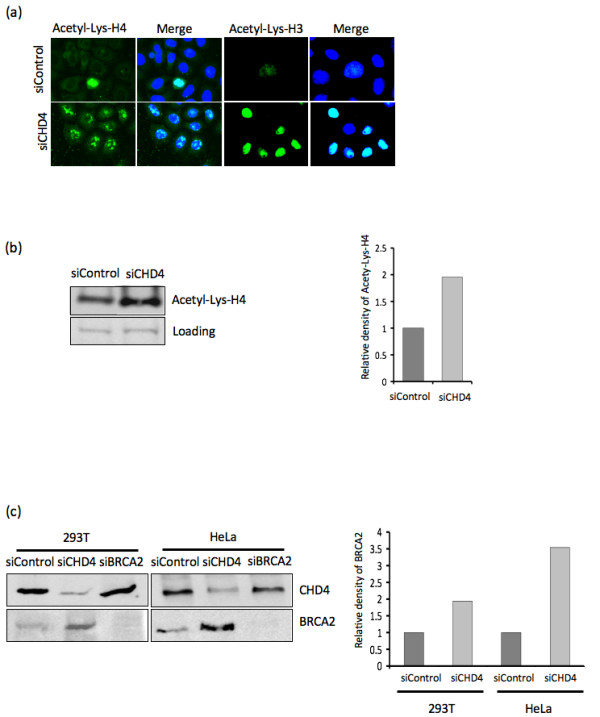
Figure 4.

CHD4 depletion leads to increased levels of euchromatin. (A) U2OS cells were mock treated (siControl) or CHD4 depleted (siCHD4), 48 hr following treatment the cells were detergent extracted to remove non-chromatin bound proteins prior to fixation. Immunofluorescence analysis for Acetylated-lysine histone H4 (Acetyl-Lys-H4) and Acetylated-lysine histone H3 (Acetyl-Lys-H3) was then performed as indicated. The merge indicates co-staining with dapi stained nuclei. (B) U2OS cells were treated with mock (siControl) or CHD4 depleted (siCHD4), 48 hr following treatment cell lysates were prepared and subjected to western blot analysis for Acetyl-Lys-H4, densitometry was used to calculate the relative protein levels. Loading indicates a non-specific band common in each lysate. (C) 293T and HeLa cells were mock treated (siControl), CHD4 depleted (siCHD4) or BRCA2 depleted (siBRCA2), 48 hr following treatment cell lysates were prepared and subjected to western blot analysis for CHD4 and BRCA2 as indicated. Densitometry was used to calculate the relative levels of each protein.
