
Fig. 1.
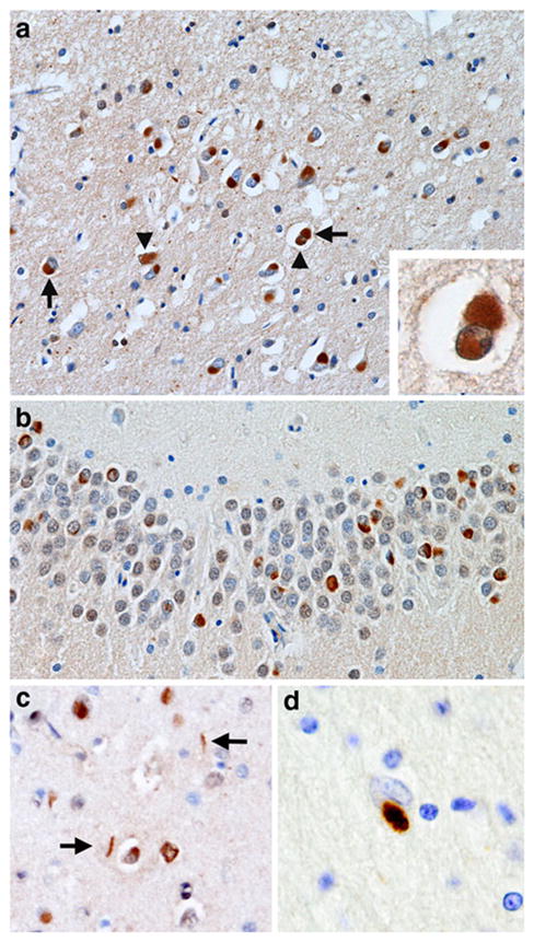
Spectrum of ‘fused in sarcoma’ (FUS) immunoreactive inclusions in neuronal intermediate filament inclusion disease (NIFID). a A neuronal cytoplasmic inclusion (NCI) (arrow) and neuronal intranuclear inclusion (NII) (arrowhead) in the superficial laminae of the frontal lobe (×200); inset shows a neuron containing both an NCI and NII; b NCI in the dentate fascia (×400); c NCI and dystrophic neurites (DN) in the frontal lobe (×200); d a glial cytoplasmic inclusion (GI) in an astrocyte in the white matter of the frontal lobe (×400); a–d FUS immunohistochemistry