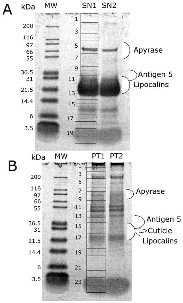
Figure 1.

One-dimensional polyacrylamide gel electrophoresis separation of the soluble (A) and insoluble (B) fractions of Dipetalogaster maxima salivary proteins. The grids indicate how the duplicated gel bands containing the salivary proteins (SN1, SN2, PT1, and PT2) were cut for tryptic digestion and mass spectrometric detection of peptides following nano reversed-phase high-performance liquid chromatography. The left of the figures indicate the molecular weight of the markers (kDa) indicated in the MW lane. For more information, see text.
