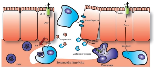
Figure 9.
Epithelial destruction and inflammation caused by E. histolytica. E. histolytica induces a flask-shaped ulcer usually spanning several villi rather than several cells as shown here. The epithelial layer is destroyed both due to pathogen factors, such as the pore forming amoebapores and extracellular matrix cleavage by cysteine proteases, as well as host inflammatory factors. E. histolytica releases PGE2 and causes a host PGE2 response that results in colonic Cl− secretion, IL-8 secretion and PMN recruitment. While PMNs can be neutralized and phagocytosed by the parasite and complement is degraded by cysteine proteases, the infection is still highly inflammatory. Macrophages have been shown to be activated by parasite DNA through TLR9 or alternately through lipophoshpopeptidoglycan activation of TLR 2 or 4. In addition to PGE2, E. histolytica also secretes serotonin, which is capable of activating Ca+ dependent Cl− secretion in the small intestine.

