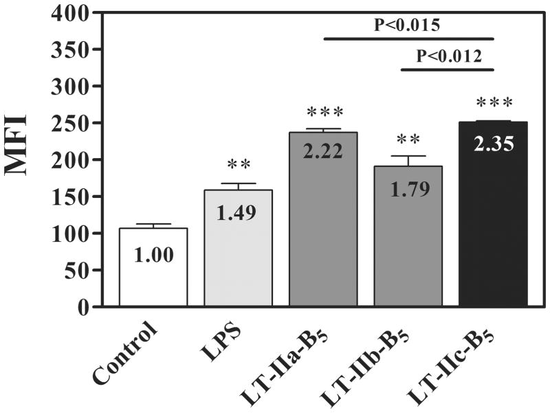
Figure 4. Uptake of FITC-OVA by BMDC is enhanced by LT-IIc-B5.
BMDC were incubated at 37°C with 5 μg/ml of LT-IIa-B5, 5 μg/ml of LT-IIb-B5, 5 μg/ml of LT-IIc-B5, 1 μg/ml of LPS, or PBS (untreated). After 10 min, FITC-OVA (0.2 mg/ml) was added to the cultures. Ten min after addition of FITC-OVA, cells were washed with ice-cold PBS, stained with an APC-conjugated anti-CD11c mAb (BioLegend) and 7-AAD (Calbiochem) and the mean fluorescent intensity (MFI) of CD11c-positive, 7-AAD-negative cells was determined using flow cytometry. Data are reported as arithmetic means; error bars denote one standard error of the mean (n = 3). Data shown represents one of two independent experiments. Fold increases (MFI levels in treated cells/MFI levels in untreated cells) for each group of cells are denoted. Key: statistically different from untreated BMDC at P < 0.005 (**) and P < 0.0001 (***); statistical differences between the values for LT-IIc-B5 & LT-IIa-B5 and LT-IIc-B5 & LT-IIb-B5 are denoted above the crossbars.

