
Figure 4.
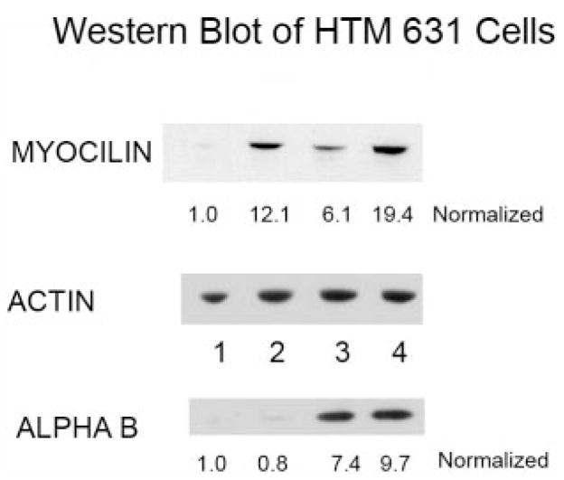
Western blot of myocilin, αB-crystallin, and actin for the homogenate of the HTM 631 cells. The actin blot was used to normalize the loading of the samples. The normalized values for each of the conditions: (1) control planar, (2) control 400-nm pitch, (3) dexamethasone-treated planar, and (4) dexamethasone treated 400-nm pitch are listed below the Western blots. In each case, the value for the control planar was set at 1.0. Although the myocilin protein levels were increased substantially in cells grown on patterned surfaces, the αB-crystallin level was similar on both planar and 400-nm pitch. However, the amount of this crystallin increased with dexamethasone treatment on both surfaces.