Figure 7.
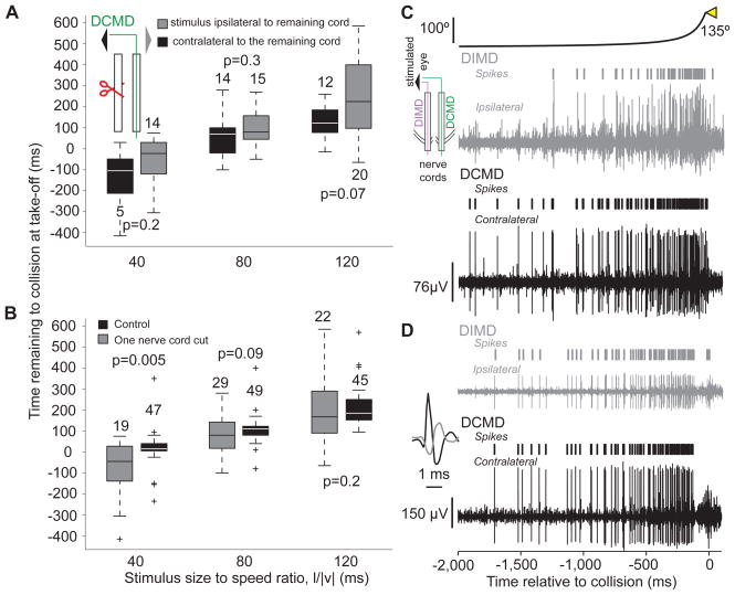
Locusts with one nerve cord sectioned still jump in response to looming stimuli and comparison between looming evoked activities in the DCMD and DIMD (see also Supp. Fig. S5). A) In animals with one sectioned nerve cord, no significant difference in the timing of take-off was observed, irrespective of the stimulated eye (box plots; nL = 6). Inset: stimulated eye and sectioning procedure (red scissors). B) The timing of take-off was significantly delayed at l/|v|=40 ms relative to control. The timing of take-off showed higher variability after cutting one nerve cord. Box plots of data pooled across trials where the stimulus was presented to the eye ipsi- and contralateral to the remaining nerve cord, since those take-off times did not show a significant difference (A). C) Looming-evoked activities in the DCMD and DIMD obtained by simultaneous recording from both nerve cords (inset). In this locust, the DCMD and DIMD spikes were not always coincident. D) Recording from a different locust in which the DCMD and DIMD spikes were coincident. Inset: example of coincident DIMD (gray) and DCMD spikes on an expanded time scale. The ipsi- and contralateral extracellular recordings are plotted on the same vertical scale.

