


Fig. 13.
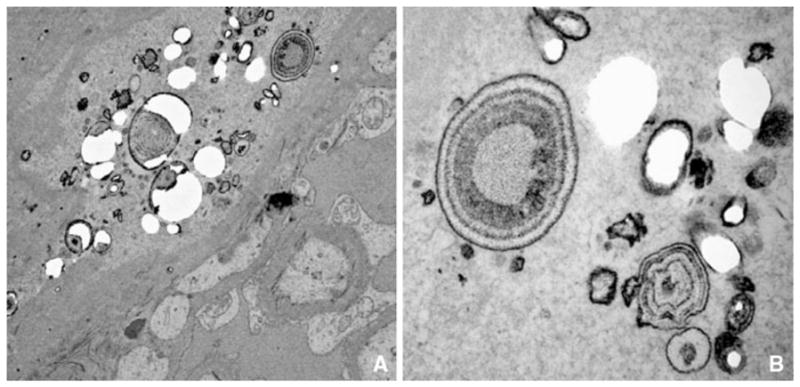
Transmission electron micrograph of interstitial deposits. At low magnification, a the plaque particles are spherical in shape and multilayered. At a higher magnification, the deposits from this THP-null mouse look identical to that seen in human idiopathic calcium oxalate stone formers (Fig. 5a)