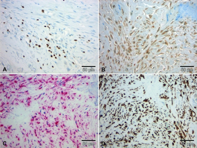
Figure 7.
Immunohistochemical staining profile of the tumor in case 1. (A) A few cells showed nuclear expression of Ki67. (B) Almost all neoplastic cells showed weak immunoreacitivity for S100. All spindle cells revealed nuclear and cytoplasmic expression of (C) vimentin and (D) smooth-muscle actin. Scale bar, 50 µm.

