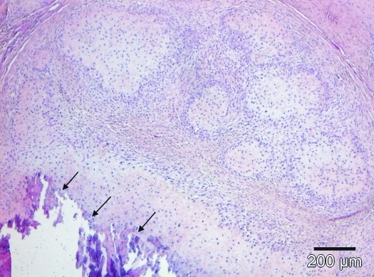
Figure 9.
The femoral tumor was composed of lobules with a poorly cellular cartilaginous matrix core surrounded by a densely cellular rim of undifferentiated polygonal mesenchymal cells. Multifocally, central lobule parts underwent dystrophic calcification (arrows). Hematoxylin and eosin stain; scale bar, 200 µm.

