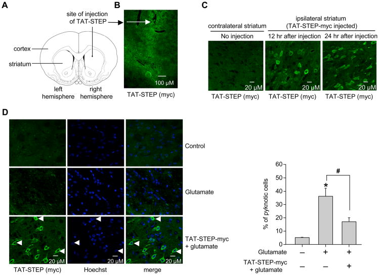
Figure 6.
TAT-STEP-myc peptide reduces glutamate-induced cell death in the striatum. (A) Schematic representation of the area of infusion of TAT-STEP-myc peptide. (B-C) TAT-STEP-myc peptide was stereotactically injected into the striatum. Coronal sections were processed for immunohistochemical analysis with anti-myc antibody at the specified time periods. (B) Low magnification (5X) and (C) high magnification (20X) images. (D) Glutamate was injected stereotactically into the striatum in the presence or absence of TAT-STEP-myc peptide. Striatal brain sections were processed for immunohistochemical analysis with anti-myc antibody and Hoechst DNA stain (left panel). Quantitative analysis of the percentage of neurons with pyknotic nuclei is represented as mean ± SEM (n ≥ 3, right panel). *Indicates significant difference from glutamate treated samples (p < 0.001).

