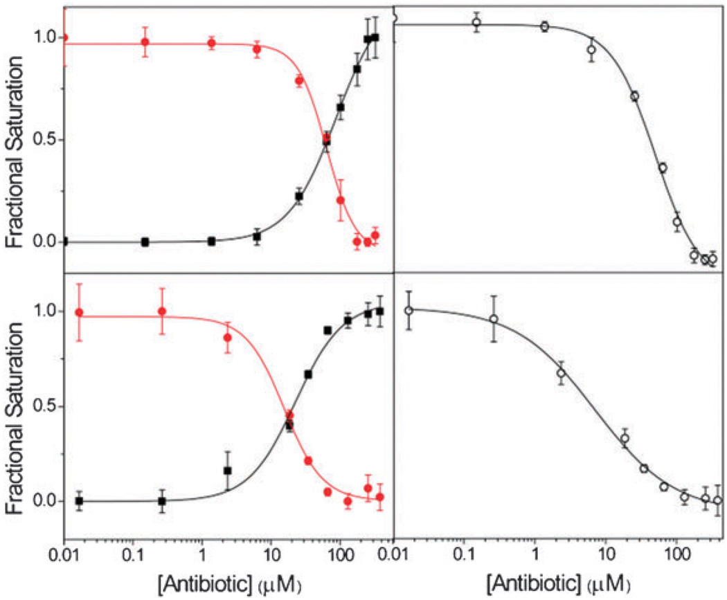
Fig. 5.
Fractional fluorescence saturation of the donor F1 (■) in the labeled 16S A-site, the emissive fluorophore F2 ( ) tagged to kanamycin A, and the emissive acceptor F3 (○) of the 18S A-site in studying the binding of: (top) negamycin; (bottom) neamine. Conditions: 16S RNA (5 × 10⁻⁷ M), 18S RNA (5 × 10⁻⁷ M), coumarin-labeled-kanamycin A (2.2 × 10⁻⁶ M), cacodylate buffer pH 7.0 (2.0 × 10⁻² M), NaCl (1.0 × 10⁻¹ M).13

