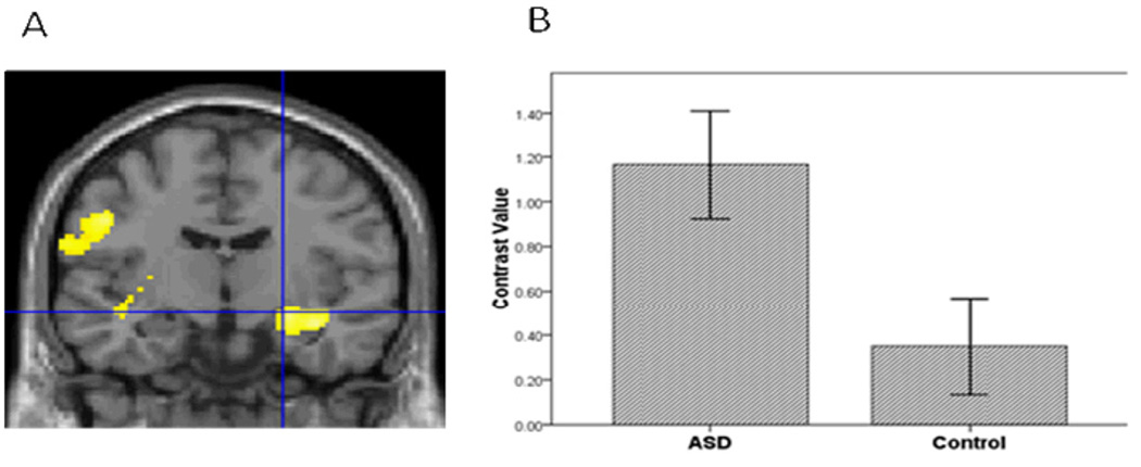
Figure 1.
A. Relative to the controls, the ASD group had greater amygdala activation in the contrast of Sad vs. Baseline. Threshold for this and subsequent figures was p=.005 for the images. B. To illustrate the activation for this and subsequent bar graphs, mean contrast values were extracted from the entire ROI within hemisphere. Contrast values represent the difference in mean activation in the ROI for a given contrast for all subjects averaged together in each group. Error bars for all figures represent standard errors of the means.

